


 膜杰作
膜杰作 Star Staining


分子别名(Synonym)
FCGR2B,C,CD32b,c,FcRII-b,c,Fc-gamma RII-b,c,Fc-gamma-RIIb,c,CD32,FCG2,IGFR2,CDw32
表达区间及表达系统(Source)
Human CD32b/c, His Tag (CDB-H5228) is expressed from human 293 cells (HEK293). It contains AA Ala 46 - Pro 217 (Accession # P31994-1). In the region Ala 46 - Pro 217, the AA sequence of Fc gamma RIIB and Fc gamma RIIC are homologus.
Predicted N-terminus: Ala 46
Request for sequence
蛋白结构(Molecular Characterization)
This protein carries a polyhistidine tag at the C-terminus.
The protein has a calculated MW of 20.4 kDa. The protein migrates as 28-31 kDa when calibrated against Star Ribbon Pre-stained Protein Marker under reducing (R) condition (SDS-PAGE) due to different glycosylation.
内毒素(Endotoxin)
Less than 1.0 EU per μg by the LAL method.
纯度(Purity)
>95% as determined by SDS-PAGE.
>90% as determined by SEC-MALS.
制剂(Formulation)
Lyophilized from 0.22 μm filtered solution in PBS, pH7.4 with trehalose as protectant.
Contact us for customized product form or formulation.
重构方法(Reconstitution)
Please see Certificate of Analysis for specific instructions.
For best performance, we strongly recommend you to follow the reconstitution protocol provided in the CoA.
存储(Storage)
For long term storage, the product should be stored at lyophilized state at -20°C or lower.
Please avoid repeated freeze-thaw cycles.
This product is stable after storage at:
- -20°C to -70°C for 12 months in lyophilized state;
- -70°C for 12 months under sterile conditions after reconstitution.
质量管理控制体系(QMS)
电泳(SDS-PAGE)
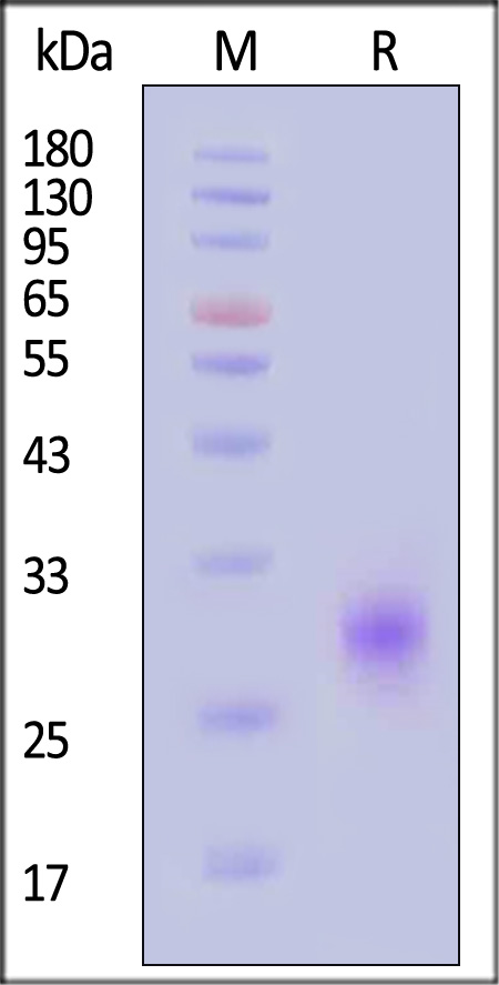
Human CD32b/c, His Tag on SDS-PAGE under reducing (R) condition. The gel was stained with Coomassie Blue. The purity of the protein is greater than 95% (With Star Ribbon Pre-stained Protein Marker).
SEC-MALS
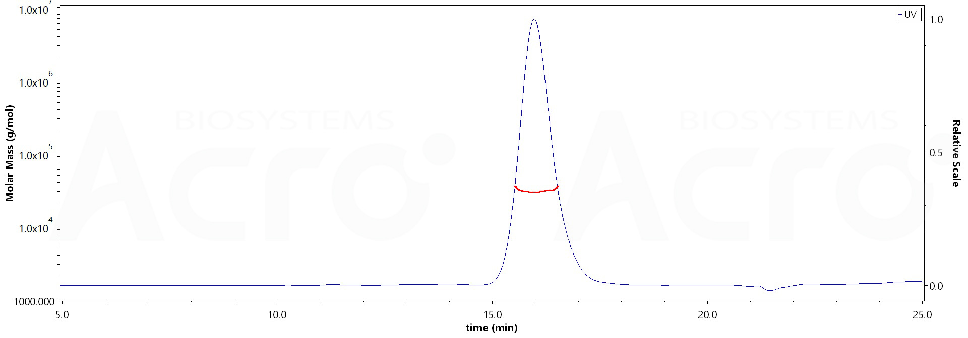
The purity of Human CD32b/c, His Tag (Cat. No. CDB-H5228) is more than 90% and the molecular weight of this protein is around 20-30 kDa verified by SEC-MALS.
Report
活性(Bioactivity)-SPR
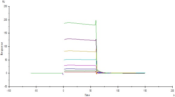
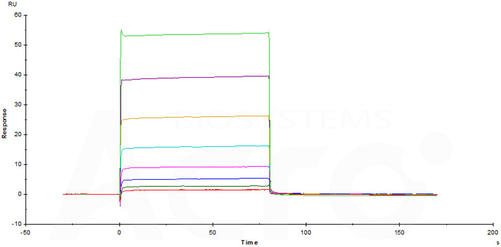
Immobilized Human CD32b/c, His Tag (Cat. No. CDB-H5228) on CM5 Chip via anti-His antibody, can bind Rituximab with an affinity constant of 15.1 μM as determined in a SPR assay (Biacore T200) (QC tested).
Protocol
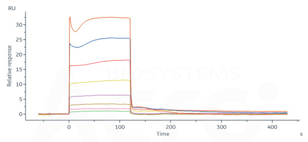
Immobilized Rituximab on CM5 Chip, can bind Human CD32b/c, His Tag (Cat. No. CDB-H5228) with an affinity constant of 5.5 μM as determined in a SPR assay (Biacore T200) (Routinely tested).
Protocol
Rituximab captured on Protein A Chip can bind Human CD32b/c, His Tag (Cat. No. CDB-H5228) with an affinity constant of 3.56 μM as determined in SPR assay (Biacore 8K) (Routinely tested).
Protocol
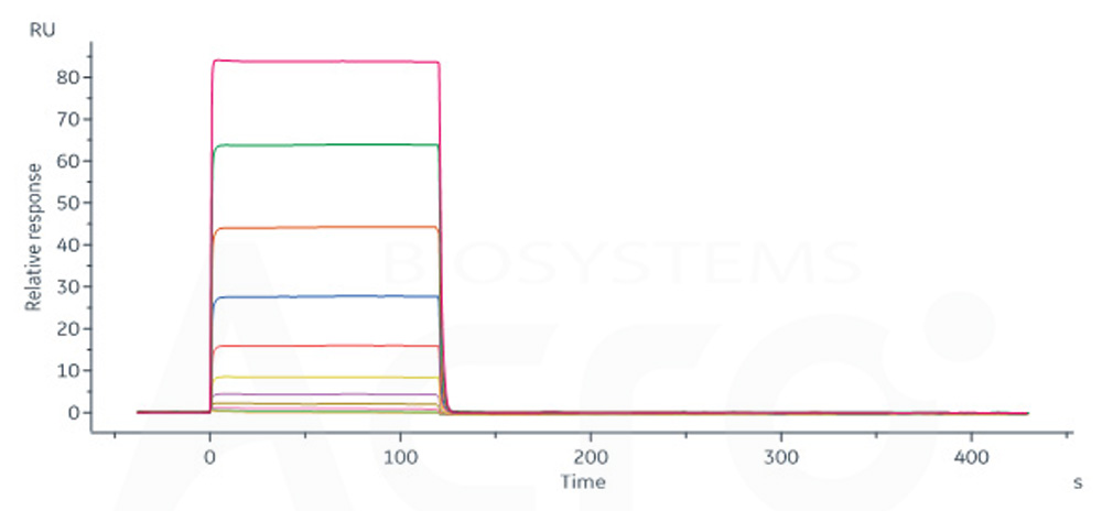
Rituximab immobilized on CM5 Chip can bind Human CD32b/c, His Tag (Cat. No. CDB-H5228) with an affinity constant of 4.09 μM as determined in SPR assay (Biacore 8K) (Routinely tested).
Protocol
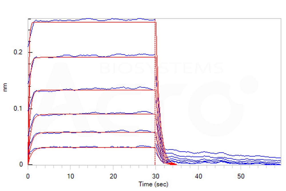
活性(Bioactivity)-BLI
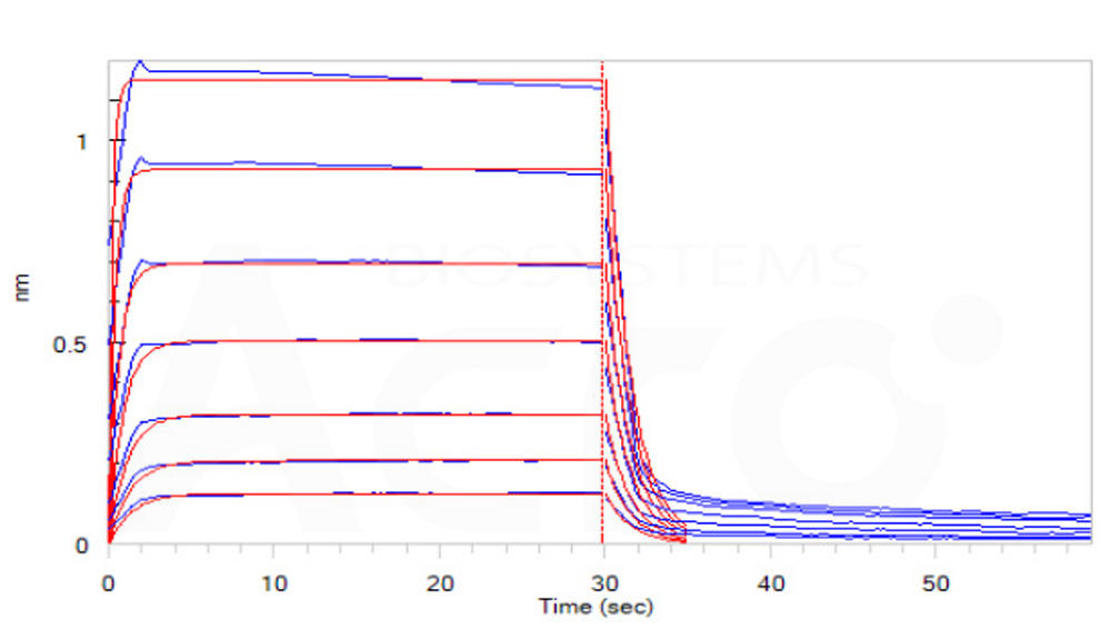
Loaded Human CD32b/c, His Tag (Cat. No. CDB-H5228) on HIS1K Biosensor, can bind Rituximab with an affinity constant of 4.30 μM as determined in BLI assay (ForteBio Octet Red96e) (Routinely tested).
Protocol
Loaded Rituximab on FAB2G Biosensor, can bind Human CD32b/c, His Tag (Cat. No. CDB-H5228) with an affinity constant of 5.4 μM as determined in BLI assay (ForteBio Octet Red96e) (Routinely tested).
Protocol
 +添加评论
+添加评论
产品推荐(Recommended Products)

背景(Background)
Receptors for the Fc region of IgG (Fc γ R) are members of the Ig superfamily that function in the activation or inhibition of immune responses. Three classes of human Fc γ Rs: RI (CD64), RII (CD32), and RIII (CD16), which generate multiple isoforms, are recognized.
There are three genes for human Fcγ RII /CD32 (A, B, and C) and one for mouse Fcγ RII B (CD32B). CD32 is a low affinity receptor for IgG. Low affinity immunoglobulin gamma Fc region receptor II-b (FCGR2B) is also known as CD32b, FCG2, IGFR2. CD32B is expressed on B cells and myeloid dendritic cells. Ligation of CD32B on B cells downregulates antibody production and may, in some circumstances, promote apoptosis. Co-ligation of CD32B on dendritic cells inhibits maturation and blocks cell activation. CD32B may also be a target for monoclonal antibody therapy for malignancies.


